File:Gray710.png
Gray710.png (400 × 452 pixels, file size: 46 KB, MIME type: image/png)
Captions
Captions
Summary
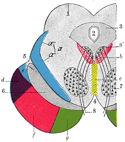
[edit]| Description | Axial section through mid-brain. (Schematic.) (Testut.) 1. Corpora quadrigemina. 2. Cerebral aqueduct. 3. Central gray stratum. 4. Interpeduncular space. 5. Sulcus lateralis. 6. Substantia nigra. 7. Red nucleus of tegmentum. 8. Oculomotor nerve, with 8’, its nucleus of origin. a. Lemniscus (in blue) with a’ the medial lemniscus and a" the lateral lemniscus. b. Medial longitudinal fasciculus. c. Raphé. d. Temporopontine fibers. e. Portion of medial lemniscus, which runs to the lentiform nucleus and insula. f. Cerebrospinal fibers. g. Frontopontine fibers. | ||||||||||||||||||||
| Plate | 710 | ||||||||||||||||||||
| Date | before 1858 | ||||||||||||||||||||
| Source |
|
||||||||||||||||||||
| Author |
|
||||||||||||||||||||
.jpg)
Book
[edit]| Henry Gray: Gray's Anatomy (20th edition)
|
|||||||||||||||||||||||
|---|---|---|---|---|---|---|---|---|---|---|---|---|---|---|---|---|---|---|---|---|---|---|---|
| Author |
|
-_Title_page.png) | |||||||||||||||||||||
| Editor |
Revised by Warren H. Lewis |
||||||||||||||||||||||
| Illustrator |
|
||||||||||||||||||||||
| Title | |||||||||||||||||||||||
| Edition |
20 |
||||||||||||||||||||||
| Publisher | |||||||||||||||||||||||
| Object type |
version, edition or translation |
||||||||||||||||||||||
| Page overview | list of all the plates | ||||||||||||||||||||||
| Language |
English |
||||||||||||||||||||||
| Publication date |
1918 |
||||||||||||||||||||||
| Place of publication |
Philadelphia / New York City |
||||||||||||||||||||||
| Source | Bartleby | ||||||||||||||||||||||

Licensing
[edit]
This image is in the public domain because it is a mere mechanical scan or photocopy of a public domain original, or – from the available evidence – is so similar to such a scan or photocopy that no copyright protection can be expected to arise. The original itself is in the public domain for the following reason:
This tag is designed for use where there may be a need to assert that any enhancements (eg brightness, contrast, colour-matching, sharpening) are in themselves insufficiently creative to generate a new copyright. It can be used where it is unknown whether any enhancements have been made, as well as when the enhancements are clear but insufficient. For known raw unenhanced scans you can use an appropriate {{PD-old}} tag instead. For usage, see Commons:When to use the PD-scan tag.  | ||||
File history
Click on a date/time to view the file as it appeared at that time.
| Date/Time | Thumbnail | Dimensions | User | Comment | |
|---|---|---|---|---|---|
| current | 19:49, 23 January 2007 | | 400 × 452 (46 KB) | Pngbot (talk | contribs) | optimized with optipng |
| 19:38, 21 January 2006 |  | 400 × 452 (73 KB) | Arcadian (talk | contribs) | {{Gray's Anatomy plate}} |
You cannot overwrite this file.
File usage on Commons
The following 2 pages use this file:
File usage on other wikis
The following other wikis use this file:
- Usage on ar.wikipedia.org
- Usage on bg.wikipedia.org
- Usage on bs.wikipedia.org
- Usage on cs.wikipedia.org
- Usage on de.wikipedia.org
- Usage on de.wikibooks.org
- Usage on en.wikipedia.org
- Cerebral aqueduct
- Medial lemniscus
- Oculomotor nucleus
- Medial longitudinal fasciculus
- Lateral lemniscus
- Periaqueductal gray
- Lentiform nucleus
- Corpora quadrigemina
- Cerebrospinal fibers
- Frontopontine fibers
- Temporopontine fibers
- Rostral interstitial nucleus of medial longitudinal fasciculus
- User:WillHHudson/Sandbox
- Corticopontine fibers
- User:Was a bee/Gray
- Usage on eo.wikipedia.org
- Usage on es.wikipedia.org
- Usage on eu.wikipedia.org
- Usage on fa.wikipedia.org
- Usage on fr.wikipedia.org
- Usage on hy.wikipedia.org
- Usage on it.wikipedia.org
- Usage on ja.wikipedia.org
- Usage on ko.wikipedia.org
View more global usage of this file.