File:Gray236.png
Original file (485 × 700 pixels, file size: 49 KB, MIME type: image/png)
Captions
Captions
Summary
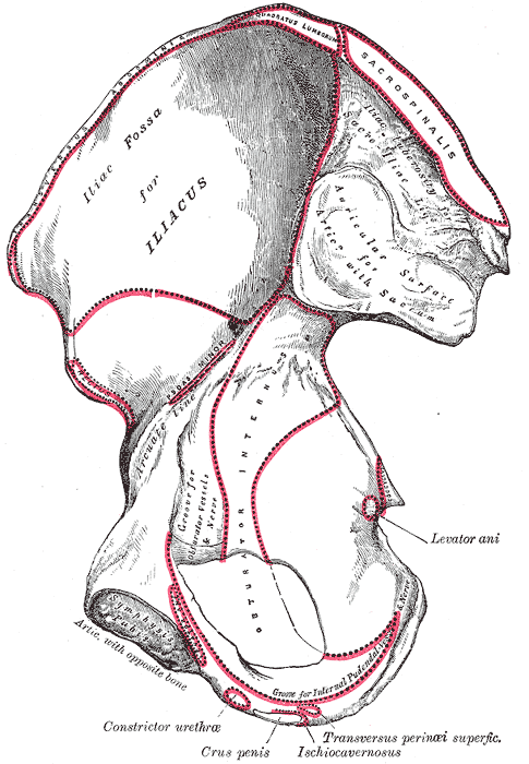
[edit]| Description | Right hip bone, internal surface. | ||||||||||||||||||||
| Plate | 236 | ||||||||||||||||||||
| Date | before 1858 | ||||||||||||||||||||
| Source |
|
||||||||||||||||||||
| Author |
|
||||||||||||||||||||
| Other versions | العربيَّة | ||||||||||||||||||||
.jpg)
Book
[edit]| Henry Gray: Gray's Anatomy (20th edition)
|
|||||||||||||||||||||||
|---|---|---|---|---|---|---|---|---|---|---|---|---|---|---|---|---|---|---|---|---|---|---|---|
| Author |
|
-_Title_page.png) | |||||||||||||||||||||
| Editor |
Revised by Warren H. Lewis |
||||||||||||||||||||||
| Illustrator |
|
||||||||||||||||||||||
| Title | |||||||||||||||||||||||
| Edition |
20 |
||||||||||||||||||||||
| Publisher | |||||||||||||||||||||||
| Object type |
version, edition or translation |
||||||||||||||||||||||
| Page overview | list of all the plates | ||||||||||||||||||||||
| Language |
English |
||||||||||||||||||||||
| Publication date |
1918 |
||||||||||||||||||||||
| Place of publication |
Philadelphia / New York City |
||||||||||||||||||||||
| Source | Bartleby | ||||||||||||||||||||||

Licensing
[edit]
This image is in the public domain because it is a mere mechanical scan or photocopy of a public domain original, or – from the available evidence – is so similar to such a scan or photocopy that no copyright protection can be expected to arise. The original itself is in the public domain for the following reason:
This tag is designed for use where there may be a need to assert that any enhancements (eg brightness, contrast, colour-matching, sharpening) are in themselves insufficiently creative to generate a new copyright. It can be used where it is unknown whether any enhancements have been made, as well as when the enhancements are clear but insufficient. For known raw unenhanced scans you can use an appropriate {{PD-old}} tag instead. For usage, see Commons:When to use the PD-scan tag.  | ||||
File history
Click on a date/time to view the file as it appeared at that time.
| Date/Time | Thumbnail | Dimensions | User | Comment | |
|---|---|---|---|---|---|
| current | 18:09, 23 January 2007 | | 485 × 700 (49 KB) | Pngbot (talk | contribs) | optimized with optipng |
| 11:32, 18 May 2006 |  | 485 × 700 (51 KB) | File Upload Bot (Magnus Manske) (talk | contribs) | {{Information| |Description= {{Gray's Anatomy plate|Right Os Innominatum, internal surface.}} |Source=Originally from [http://en.wikipedia.org en.wikipedia]; description page is (was) here * 08:42, 22 September 2003 [[:en:User:M |
You cannot overwrite this file.
File usage on Commons
The following 3 pages use this file:
File usage on other wikis
The following other wikis use this file:
- Usage on ar.wikipedia.org
- Usage on az.wikipedia.org
- Usage on bg.wikipedia.org
- Usage on bn.wikipedia.org
- Usage on bs.wikipedia.org
- Usage on ca.wikipedia.org
- Usage on ckb.wikipedia.org
- Usage on cs.wikipedia.org
- Usage on de.wikipedia.org
- Usage on el.wikipedia.org
- Usage on en.wikipedia.org
- Levator ani
- Ischiocavernosus muscle
- Internal pudendal artery
- Psoas minor muscle
- External sphincter muscle of male urethra
- Iliacus muscle
- Anterior superior iliac spine
- Ilium (bone)
- Obturator nerve
- Ischium
- Pubis (bone)
- Erector spinae muscles
- Obturator artery
- Ischial tuberosity
- Wing of ilium
- Iliac fossa
- Iliac tuberosity
- Iliac tubercle
- Hip bone
- User:Was a bee/Gray
- Vaginal support structures
- Usage on es.wikipedia.org
View more global usage of this file.