File:Gray193.png

Original file (719 × 1,057 pixels, file size: 150 KB, MIME type: image/png)
Captions
Captions
Summary
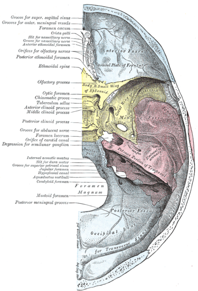
[edit]| Description | Base of the en:skull. Inner or cerebral surface. | ||||||||||||||||||||
| Plate | 193 | ||||||||||||||||||||
| Date | before 1858 | ||||||||||||||||||||
| Source |
|
||||||||||||||||||||
| Author |
|
||||||||||||||||||||
| Other versions | Derivative works of this file: Gray193 notext.png | ||||||||||||||||||||
.jpg)
Book
[edit]| Henry Gray: Gray's Anatomy (20th edition)
|
|||||||||||||||||||||||
|---|---|---|---|---|---|---|---|---|---|---|---|---|---|---|---|---|---|---|---|---|---|---|---|
| Author |
|
-_Title_page.png) | |||||||||||||||||||||
| Editor |
Revised by Warren H. Lewis |
||||||||||||||||||||||
| Illustrator |
|
||||||||||||||||||||||
| Title | |||||||||||||||||||||||
| Edition |
20 |
||||||||||||||||||||||
| Publisher | |||||||||||||||||||||||
| Object type |
version, edition or translation |
||||||||||||||||||||||
| Page overview | list of all the plates | ||||||||||||||||||||||
| Language |
English |
||||||||||||||||||||||
| Publication date |
1918 |
||||||||||||||||||||||
| Place of publication |
Philadelphia / New York City |
||||||||||||||||||||||
| Source | Bartleby | ||||||||||||||||||||||

Licensing
[edit]
This image is in the public domain because it is a mere mechanical scan or photocopy of a public domain original, or – from the available evidence – is so similar to such a scan or photocopy that no copyright protection can be expected to arise. The original itself is in the public domain for the following reason:
This tag is designed for use where there may be a need to assert that any enhancements (eg brightness, contrast, colour-matching, sharpening) are in themselves insufficiently creative to generate a new copyright. It can be used where it is unknown whether any enhancements have been made, as well as when the enhancements are clear but insufficient. For known raw unenhanced scans you can use an appropriate {{PD-old}} tag instead. For usage, see Commons:When to use the PD-scan tag.  | ||||
File history
Click on a date/time to view the file as it appeared at that time.
| Date/Time | Thumbnail | Dimensions | User | Comment | |
|---|---|---|---|---|---|
| current | 11:27, 18 May 2006 | | 719 × 1,057 (150 KB) | File Upload Bot (Magnus Manske) (talk | contribs) | {{Information| |Description= {{Gray's Anatomy plate|Base of the en:skull. Inner or cerebral surface.}} |Source=Originally from [http://en.wikipedia.org en.wikipedia]; description page is (was) here * 12:02, 19 September 20 |
You cannot overwrite this file.
File usage on Commons
The following 5 pages use this file:
File usage on other wikis
The following other wikis use this file:
- Usage on ar.wikipedia.org
- عظم صدغي
- عظم وتدي
- جيب سهمي علوي
- ثقوب شمية
- ناتئ وداجي
- ثقبة عوراء للعظم الجبهي
- درز وتدي غربالي
- درز وتدي صدفي
- ثقبة بيضوية (جمجمة)
- حفرة القحف الأمامية
- حفرة القحف المتوسطة
- تلم تصالبي
- قاعدة الجمجمة
- ظهر السرج
- ناتئ سريري أوسط
- ناتئ سريري خلفي
- ناتئ سريري أمامي
- حفرة نخامية
- حديبة السرج التركي
- قناة بصرية
- فتحة السمع الداخلية
- لسين العظم الوتدي
- نفق تحت اللسان
- قائمة ثقوب جسم الإنسان
- شريان سحائي خلفي
- شوكة غربالية
- حديبة وداجية
- درز وتدي جبهي
- درز جبهي غربالي
- ثقبة وداجية
- درز وتدي صخري
- ثقبة ممزقة
- Usage on az.wikipedia.org
- Usage on bg.wikipedia.org
- Usage on bs.wikipedia.org
- Usage on ca.wikipedia.org
- Usage on ckb.wikipedia.org
- Usage on da.wikipedia.org
- Usage on de.wikipedia.org
- Usage on en.wikipedia.org
View more global usage of this file.
Metadata
- Gray's Anatomy plates of bones
- Anatomical plates and drawings of the human skull
- Human foramen magnum
- Human frontal bones
- Human occipital bones
- Human temporal bones
- Human sphenoid bone
- Human ethmoid bone
- Sphenosquamosal sutures
- Jugular foramen
- Internal occipital protuberance
- Groove for transverse sinus
- Internal occipital crest
- Cruciform eminence
- Sella turcica
- Hypophysial fossa
- Foramen spinosum
- Petrous part of the temporal bone
- Internal acoustic meatus
- Anterior cranial fossa
- Middle cranial fossa
- Cranial base
- Superior sagittal sinus
- Optic canal
- Internal surface of cranial base
- Human occipital bone with bony landmarks labeled
- Human cranial base with bony landmarks labeled